WELCOME TO EPme.me!!!

Instagram Gallery
Recent Blog/Vlog posts
Learn from a great case study “VT Or Not...?” where we learn the finer sensitivities of analyzing arrhythmia episodes in devices.
Learn from a great case study “The Tachycardia That’s Too Slow To Detect” where we learn the finer sensitivities of using home monitoring in cardiac devices with cardiomyopathy patients.
Learn about the “Using ILR (Insertable Loop Recorder) For Arrhythmia Diagnosis” in cardiac electrophysiology.
Learn about the “Argument For Sub-Specializing” in medicine (or any field), with a stress on cardiac electrophysiology.
Overview - Recent Posts
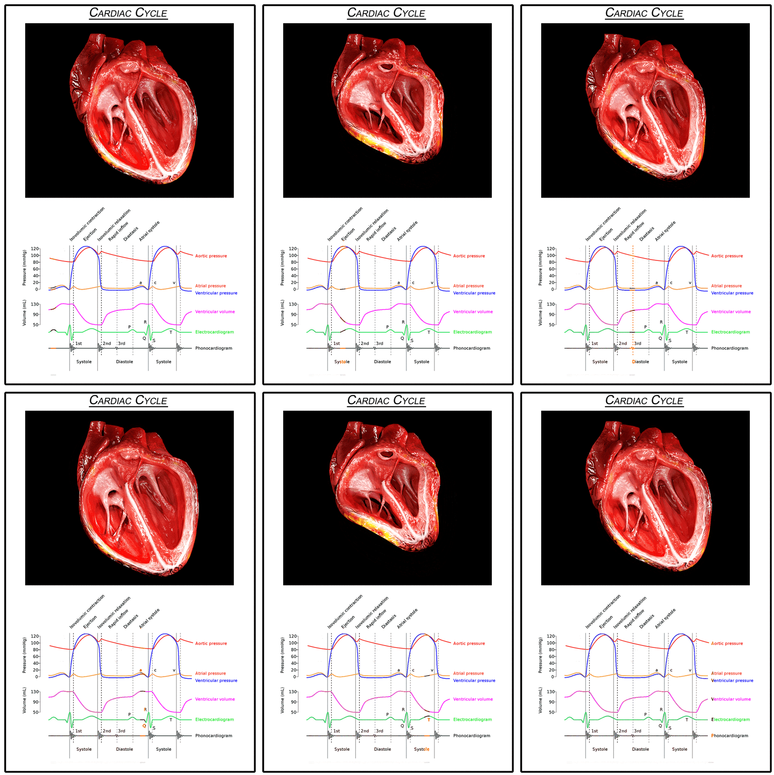
Blood flow in the heart is done by the contraction of the heart muscle.
The cardiac cycle is divided into two periods:
Diastole - The ventricle muscle is relaxed
Systole - The ventricle muscle is contracted
The circulation of blood can be divided into two cycles: the Systemic Cycle and the Pulmonary Cycle.
The heart consists of four chambers:
Right and Left Atrium, and the Right and Left Ventricles.
The Atrium is connected to Ventricles via valves and the right and left sides are separated by a septum.
The heart consists of four key valves:
- Those between the Atrium and the Ventricles - On the right side the Tricuspid Valve (3 cusps), and on the left side Mitral Valve (2 cusps).
- Those between the Ventricles and out of the heart, commonly known as the Semilunar Valves (3 cusps) - On the right side the Pulmonary Valve and on the left side Aortic Valve.
The heart consists of four chambers:
Right and Left Atrium, and the Right and Left Ventricles.
The Atrium is connected to Ventricles via valves and the right and left sides are separated by a septum.
The heart muscle itself is composed of different layers, which can be divided into three main layers (from outside to inside):
- Pericardium
- Myocardium
- Endocardial Layer (Endocardium)


Learn from a great case study “Non-Invasive EPS – Fact or Fiction?” where we learn using CRM devices for arrhythmia diagnosis.